-
Giving you the best vision
-
-

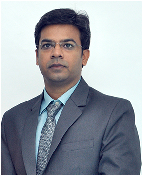
Dr. Kalyan Chothe
MBBS, Rural Medical College, Loni. (Pune University) ; F.R.C.S (Ophth), Royal College of Physicians & Surgeons, Glasgow, England.
Dr. Kalyan Chothe, F.R.C.S (Ophth) from Royal College of Physicians and Surgeons in Glasgow, England, has been serving patients with super-specialty eye care consultations and cataract, glaucoma, retinal, and other surgeries for over 17 years, emerging his professional career in 2005.
Solutions to Medical Problems
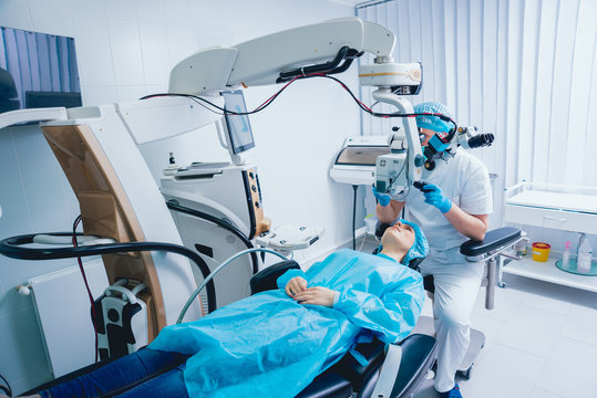
We merge two services consulting and brilliant client Services for the patient healthcare. Used latest technology in hospital.

CATARACT
A cataract is a dense, cloudy area that forms in the eye's lens, causing symptoms such as blurry vision, double vision, difficulty driving at night.
LASIK & REFRACTIVE SURGERY
LASIK surgury is performed to minimise or eliminate the need for glasses or corrective lenses. It is most prevalent type of refractive surgery.
GLAUCOMA
A glaucoma is a group of eye diseases that can cause vision loss and blindness by damaging the optic nerve in the back of the eye. It usually.
SQUINT
A squint is a condition in which the eyes do not properly align. One eye moves inward, upward, downward, or outward, while the other.
CONTACT LENSES
Sports and other physical activities benefit greatly from contact lenses. Many people use them as fashion because they are now available.
UVEITIS
Sports and other physical activities benefit greatly from contact lenses. Many people use them as fashion because they are now available.
Solutions to Medical Problems
Pediatric Ophthalmology Squint surgery
White star ICE phaco stitchless cataract surgery.
Refractive cataract surgery with imported aspheric, multifocal, toric, accommodative lenses.
LASIK for spectacle number removal.
Refractive cataract surgery.
Computerised eye checkup.
Children’s eye testing.
Contact lenses.
Retina glaucoma treatment.
Squint surgery.
Laser for diabetic retinopath

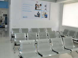
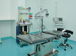
Facilities
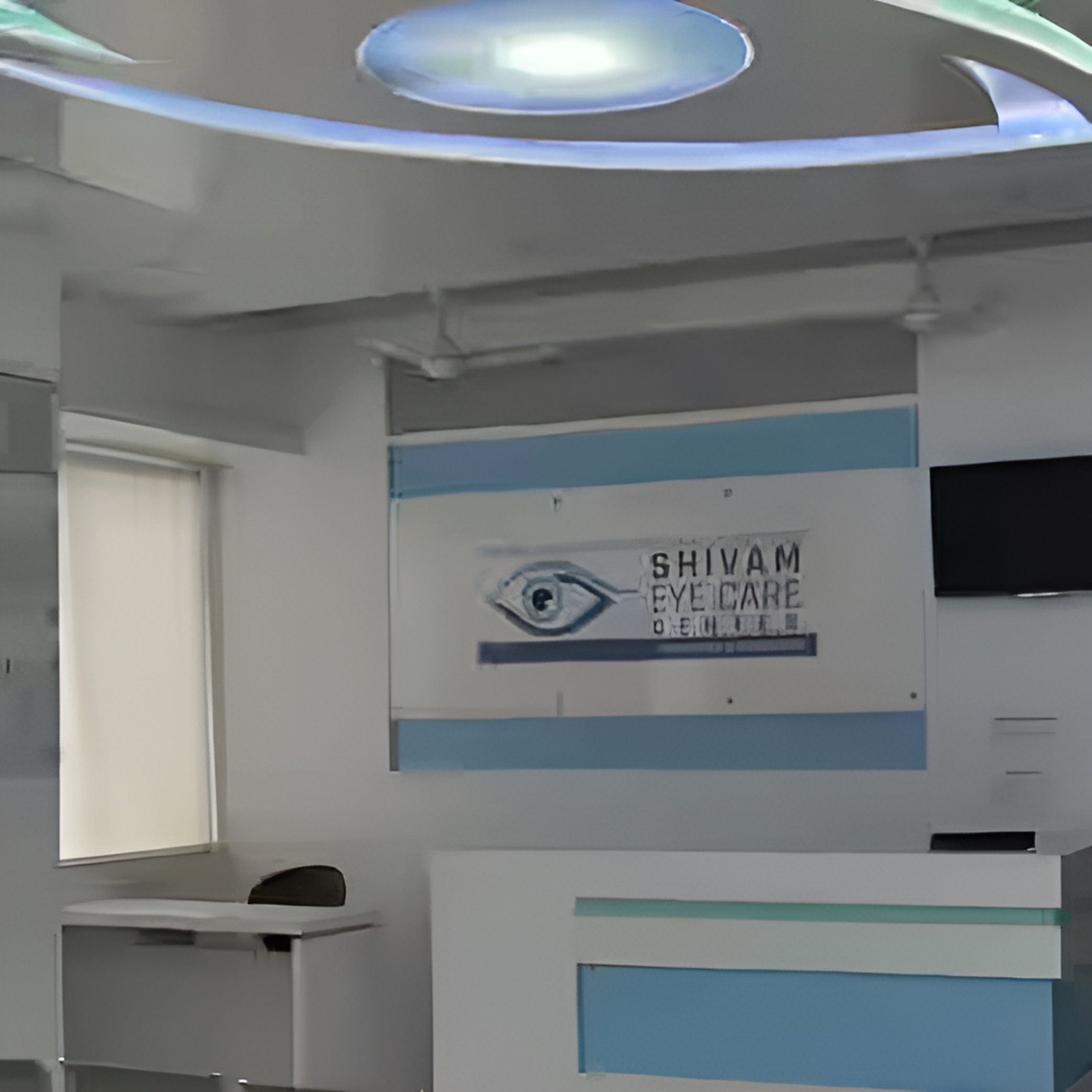
Making availability of Modern infrastructure and technologies in Maharashtra, has made Shivam Eye Care Center an icon of dynamic progress in ophthalmology. Outstanding patient outcomes have elevated it to key referral nodes in ophthalmic care community today.


What Patient say about us
Aliquam a augue suscipit, luctus neque purus ipsum neque dolor primis libero at tempus, blandit posuere ligula varius congue cursus porta feugiat
Etiam sapien sem at sagittis congue an augue massa varius egestas a suscipitEtiam sapien sem at sagittis congue an augue massa varius egestas a suscipit
Etiam sapien sem at sagittis congue an augue massa varius egestas a suscipitEtiam sapien sem at sagittis congue an augue massa varius egestas a suscipit
Etiam sapien sem at sagittis congue an augue massa varius egestas a suscipitEtiam sapien sem at sagittis congue an augue massa varius egestas a suscipit
Etiam sapien sem at sagittis congue an augue massa varius egestas a suscipitEtiam sapien sem at sagittis congue an augue massa varius egestas a suscipit
Etiam sapien sem at sagittis congue an augue massa varius egestas a suscipitEtiam sapien sem at sagittis congue an augue massa varius egestas a suscipit
Etiam sapien sem at sagittis congue an augue massa varius egestas a suscipitEtiam sapien sem at sagittis congue an augue massa varius egestas a suscipit
Etiam sapien sem at sagittis congue an augue massa varius egestas a suscipitEtiam sapien sem at sagittis congue an augue massa varius egestas a suscipit